Start with what you want to see. Explore microscope layouts, learn the controls and optics, document photographs and portable inspections, or choose a research imaging method. Two historical reports remain clearly dated below.
Getting started: choose, understand, and use
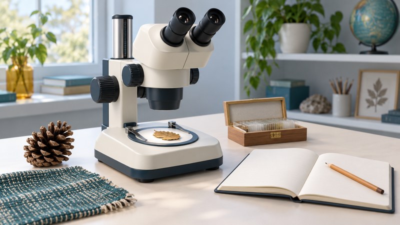
Microscopes for Kids: Choose a First Scope and Start Exploring
Match a microscope to the things a child wants to see, plan a manageable first session, and keep a useful observation record.
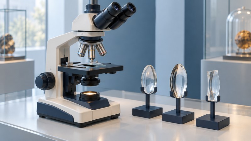
Compound Light Microscope Diagram: Follow the Light and Image
Trace the light path through a compound microscope, distinguish optical components, and calculate viewing magnification.
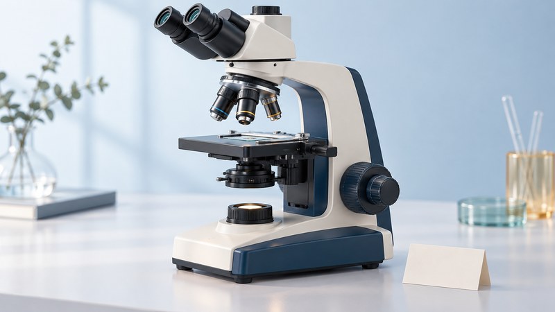
Microscope Parts Diagram: Controls, Functions, and Troubleshooting
Identify the parts of an upright compound microscope, learn what its controls change, and work through common viewing problems.
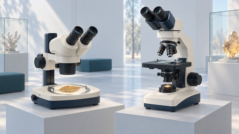
Microscope Types in Pictures: Recognize the Layout and Purpose
Use labeled schematics to recognize stereo, upright compound, inverted and digital inspection microscopes, then check the specifications a picture cannot show.
Photography, cameras, and portable inspection
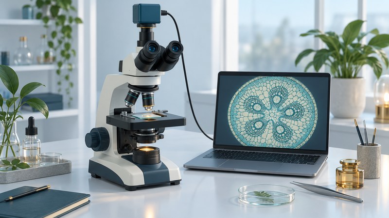
Microscope Photography: Focus, Exposure, and Accurate Scale Bars
Capture clear photomicrographs, diagnose common image problems, calibrate a scale bar, and preserve an honest record of your work.
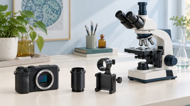
Microscope Camera Adapters: Check Fit, Optics, and Software
Compare phone, interchangeable-lens camera and dedicated microscope camera connections, then verify the complete imaging chain.
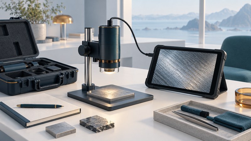
Portable Digital Microscopes: Plan a Useful Surface Inspection
Choose field of view and working distance, stabilize a portable microscope, calibrate measurements, and document which surfaces you inspected.
Choose and interpret research imaging
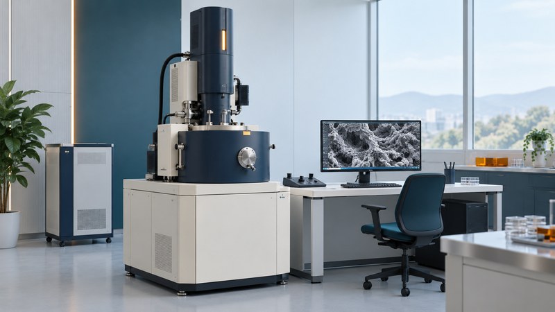
Electron Microscopes: SEM, TEM, and STEM Explained
Match an electron microscopy question to SEM, TEM or STEM, understand preparation and contrast, and prepare a useful facility consultation.
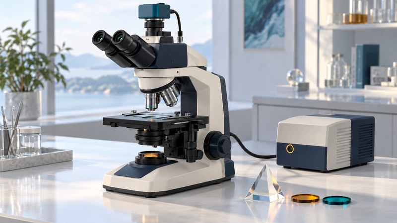
Advanced Light Microscopy: Choose the Contrast Your Question Needs
Compare brightfield, phase contrast, DIC, fluorescence and confocal imaging, then plan a specimen-specific demonstration with an imaging facility.
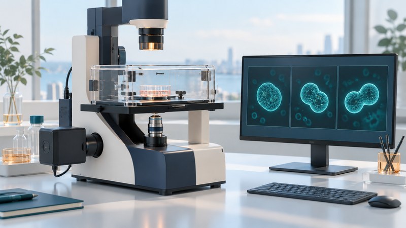
Real-Time Microscopy: Plan a Reliable Time Series
Separate preview speed from recorded timing, budget exposures and storage, and check focus drift, light burden and image-sequence artifacts.
Historical instruments and research
Read these pages as dated reporting. Product availability, prices, software support and performance claims require current verification.
- QX3 Intel Microscope — Legacy product description with Windows 98/Me/2000-era requirements; present-day compatibility is not established.
- Laser X-Ray Microscope — 2003 research report on laser-generated soft X-rays and prospective biological imaging.