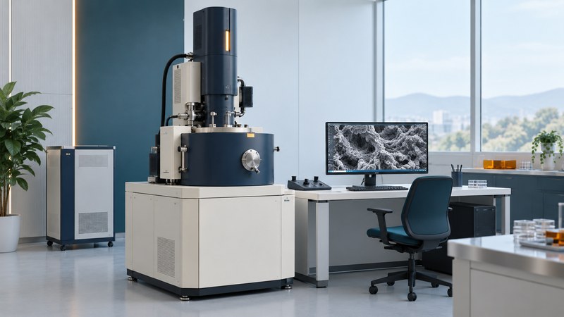
Updated October 9, 2026
Electron microscopes use electron beams and detectors to investigate structures that can be difficult to resolve with ordinary light microscopy. Start with the information you need: surface form, a thin specimen’s internal structure, or a signal associated with composition or crystal structure. That question, together with specimen preparation, points toward SEM, TEM or STEM.
Match the question to a method
| Approach | How the image is made | A useful starting question |
|---|---|---|
| SEM: scanning electron microscopy | A focused beam scans the specimen; detectors collect signals generated by its interaction with the material. | What does an exposed surface look like, and where do regions differ in the selected signal? |
| TEM: transmission electron microscopy | Electron optics form an image using electrons transmitted through a sufficiently thin specimen. | What fine structure is present in this prepared section or thin material? |
| STEM: scanning transmission electron microscopy | A small probe scans a thin specimen; detectors below it record transmitted or scattered electrons at each position. | How does structure or a chosen analytical signal vary across this thin specimen? |
These are starting distinctions, and modern instruments can combine modes. JEOL explains the basic SEM signal pathways, TEM image formation and STEM scanning and detector geometry. The same specimen can look different when the detector or imaging conditions change.
For example, secondary-electron imaging often emphasizes surface form. Backscattered-electron contrast can reveal compositional differences, but geometry and crystal orientation also affect it. Elemental analysis such as EDS records characteristic X-rays and requires its own acquisition and interpretation. Ask which signal supports each proposed conclusion.
Treat preparation as part of the evidence
An electron-microscopy session begins before the specimen enters the instrument. The facility needs to know its composition, size, substrate, moisture, conductivity, beam sensitivity and handling restrictions. A coating, polished cross-section, thin section or focused-ion-beam lamella may help answer the question while also changing what is available to observe.
Preparation is highly specimen-dependent. Ordinary high-vacuum imaging, low-vacuum modes, cryogenic workflows and specialized holders have different requirements. Agree on the method with the facility before drying, coating, cutting or sending a sample. Report hazardous constituents and follow the facility’s submission instructions.
JEOL’s 3D NAND preparation example demonstrates how a FIB-SEM can target thin TEM specimens in different directions. The lesson for a new project is to specify the location and orientation of interest before preparation: a beautiful section taken elsewhere may leave the original question unanswered.
Read a micrograph with its acquisition record
- Locate the image. Connect the detailed field to an overview, specimen identifier and preparation orientation. A selected field needs context before it can represent a larger sample.
- Identify the contrast. Read the method, detector and settings. Brightness can reflect several physical effects; an element name needs supporting analytical evidence.
- Read scale and processing. Use the calibrated scale bar and retained metadata. Screen size or printed magnification changes when a picture is resized. Describe false color, filtering, cropping and composites.
- Check competing explanations. Ask whether charging, contamination, drift, preparation damage or beam-induced change could explain a feature. Compare repeat fields or conditions with the microscopist.
- State the observation and inference separately. “A dark band is visible at this interface” is an observation. Assigning that band to a material, void or failure mechanism takes additional evidence.
A conventional SEM micrograph is a two-dimensional signal map even when shading gives a strong impression of depth. Quantitative height or three-dimensional structure requires an appropriate additional method. Likewise, a stated instrument resolution describes specified conditions; useful detail in your specimen also depends on preparation, stability, signal and sampling.
JEOL’s Scanning Electron Microscope A to Z (PDF) provides a fuller explanation of image formation, specimen preparation and imaging difficulties. Use it to frame questions for the facility, whose operating procedures govern the actual session.
Prepare a facility consultation
Bring one sentence stating the decision you want the images to support, an overview photo, an estimated feature size with units, and a list of available samples and controls. Identify which changes to the specimen are acceptable and whether it must be returned intact.
Download the electron-microscopy facility brief (TXT) to record the agreed preparation, requested signals, sampling plan, expected deliverables and interpretation responsibilities. Ask for original data, acquisition metadata and an explanation of any analysis beyond imaging.
If the question is about live behavior or fluorescent labeling, first compare the possibilities in advanced light microscopy. Technique choice follows the evidence needed, not a single magnification ranking.
Explore the Microscopes library. Sources are linked beside the explanations they support.