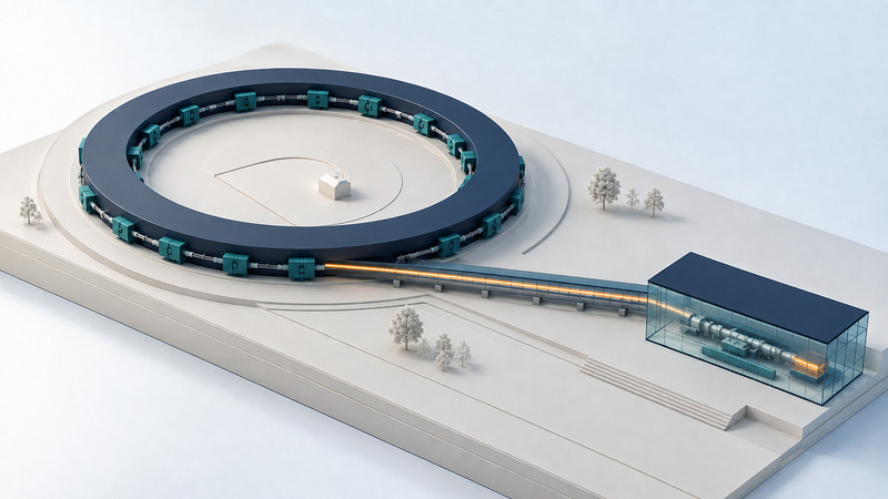
A particle accelerator can be an exceptionally useful source of light. At a synchrotron light source, energetic electrons produce radiation that scientists direct onto samples. The experiment measures what happens to those photons, using scattering, absorption, or imaging to investigate materials, chemistry, and biology.
Follow the energy from electrons to a sample
Start with charged particles. Electric fields transfer energy to them; magnetic fields steer and focus the beam. Radiofrequency cavities provide electric fields timed to the passing bunches. The particles travel through a vacuum system to limit collisions with gas molecules. CERN's explanation of accelerator operation separates these jobs of accelerating, steering, and focusing.
For a storage-ring light source, an injector system supplies electrons to a ring where they circulate repeatedly. They travel extremely close to the speed of light, but electrons have mass and do not reach the speed of light in vacuum. Increasing their energy at these speeds is not the same as increasing their speed by a comparable percentage.
When relativistic electrons bend, they emit synchrotron radiation strongly concentrated near their instantaneous direction of travel. Bending magnets therefore serve as light-source locations. In suitable straight sections, arrays of alternating magnets called undulators make electrons follow a small oscillating path and produce intense, structured radiation. SLAC's synchrotron light-source explainer describes this route from circulating electrons to useful X-rays.
Radiated energy has to be replenished. RF cavities help maintain the stored beam's energy; the ring is not an engine that produces light without an energy supply. CERN describes this compensating role of radiofrequency cavities. Meanwhile, the magnets and control systems maintain the conditions needed for a usable beam.
The emitted photons travel into a beamline, a dedicated route with optics, diagnostics, and an experimental station. Depending on the instrument, mirrors focus or redirect the light and a monochromator selects a narrow wavelength or energy range. For a concrete example, Diamond describes the mirrors and monochromator in its B18 optical layout. A detector records an interaction with the sample. The useful scientific measurement occurs at that station; the storage ring supplies the light rather than directly producing a finished molecular structure.
A synchrotron is not the same experiment as a collider
On a small screen, scroll the table sideways to read all columns.
| Facility | What happens to the beam | Typical scientific use |
|---|---|---|
| Storage-ring light source | Electrons circulate; emitted photons feed specialized beamlines. | Diffraction, absorption spectroscopy, scattering, and imaging of samples. |
| Particle collider | Opposing particle beams meet at collision points surrounded by detectors. | Study collision products and fundamental particle interactions. |
| X-ray free-electron laser (XFEL) | Accelerated electron bunches interact with their radiation in a long undulator to generate coherent X-ray pulses. | Probe very fast processes and demanding structural experiments with short, intense pulses. |
Both synchrotron light sources and XFELs can use undulators, but the laser-producing interaction in an XFEL is a key distinction. At facilities such as LCLS, electron bunches pass through a linear accelerator and an undulator; the X-rays proceed to experiments and the electrons are directed to a beam dump. SLAC explains the mechanism in its X-ray free-electron laser guide. Short pulses can help resolve rapid changes, but intense exposures can also damage samples.
A collider is organized around particle collisions and their products, rather than using an external sample as the object illuminated by a photon beam. CERN's accelerator overview provides that particle-physics context. The shared word “accelerator” describes part of the machinery, not a single scientific purpose.
No facility type wins every comparison. The relevant questions include photon energy, flux delivered to the sample, beam size, energy resolution, pulse structure, coherence, detector performance, and sample environment. Even a very bright source may not have the particular instrument a project requires.
Example: does a powder change structure when heated?
Hypothetical experiment: a research group wants to know whether a crystalline powder changes phase during a controlled heating cycle. This is a proposed workflow, not a report of an observed material or a claim that a particular beamline accepts this setup.
- Define the evidence needed. Decide which changes in diffraction peaks would support a phase change and which alternative explanations must be checked.
- Choose a compatible instrument. Discuss the sample holder, temperature range, atmosphere, X-ray energy, acquisition time, and anticipated signal with a beamline scientist.
- Collect comparison measurements. Measure appropriate standards and an empty holder or background where the method requires them. Preserve the conditions for each.
- Synchronize the records. Pair each diffraction frame with time and sample-environment measurements. A heater's setpoint is not necessarily the powder's temperature.
- Test the interpretation. Compare repeated exposures, relevant controls, and heating or cooling behavior. Consider sample motion, preferred orientation, beam-induced change, and calibration drift.
New peaks would motivate checking a new phase model. A small shift of existing peaks might reflect changing lattice dimensions, but an angular calibration problem could also shift peaks. A disappearing peak can result from something other than a vanished phase. The X-ray diffraction guide explains how to separate position, intensity, and width before drawing a conclusion.
A useful experiment therefore produces more than a sequence of attractive plots. It produces raw detector data, calibration information, acquisition settings, environmental records, and a documented analysis. The reproducible research workflow helps keep those pieces connected.
Start with the science and the beamline team
At Stanford's SSRL, SPEAR3 is the storage-ring source behind a range of X-ray experiments. Researchers seek access through the facility's proposal and review process. SSRL's user facility access policy describes general and partner access, institutional user agreements, and non-proprietary versus proprietary arrangements. Check the current facility instructions for submission dates and requirements.
Before writing a proposal, explain why the required measurement needs that instrument, what preliminary evidence supports feasibility, how samples will be handled, and how much useful data the requested time should produce. Ask about sample changes under exposure and plan a way to detect them. Access, safety review, and instrument compatibility are practical parts of experimental design.
The accelerator makes a difficult measurement possible. Controls, calibration, and analysis determine whether that measurement answers the question. Treat a striking image or a high count rate as the beginning of interpretation, not as proof on its own.
Related resources
- Read a diffraction pattern and its limitations
- Keep sample-environment measurements interpretable
- Preserve the route from raw data to result
- Explore all science resources
Researched and updated September 6, 2026. Consult the linked primary sources for methods, evidence, and limitations.