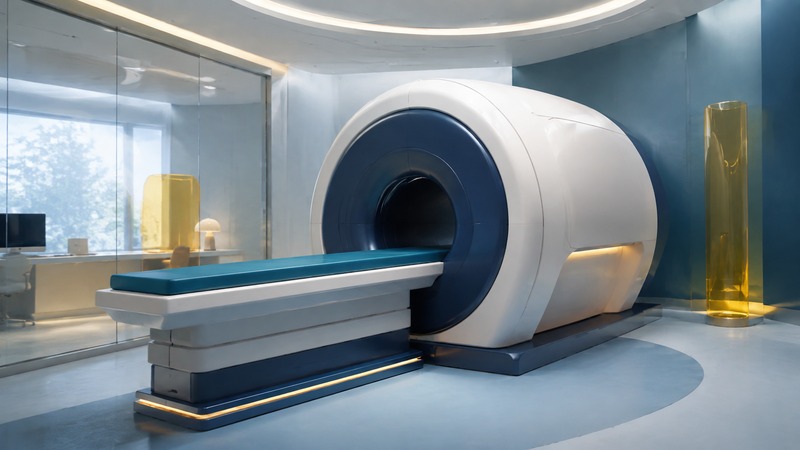
A conventional MRI machine looks like a large cylinder with a tunnel through its center. A padded table moves the part of your body being examined into the scanner. Knowing that basic layout can make the appointment easier to picture, but the shape alone does not tell you which examination is appropriate.
MRI uses magnetic fields and radiofrequency energy to produce images; it does not use the ionizing radiation used in x-rays and CT. The FDA’s MRI benefits and risks guide explains why the magnetic environment still requires careful safety screening.
Closed, wide-bore, open, and upright
On a small screen, scroll the table sideways to read all columns.
| Description | What you may see | Question for the imaging center |
|---|---|---|
| Conventional cylindrical MRI | A tunnel within a substantial magnet housing and a table aligned with it. | Where will my head and the area being examined be positioned? |
| Wide-bore MRI | A cylindrical scanner with a larger opening. | What opening and table limits apply at your facility? |
| Open MRI | A design with more open space around the patient, depending on the machine. | Can this scanner perform the exact ordered examination? |
| Upright or positional MRI | A specialized system that can image in seated, standing, or other supported positions. | Is that position needed to answer my clinician’s specific question? |
RadiologyInfo’s MRI preparation guide, produced by the American College of Radiology and Radiological Society of North America, describes conventional, wider, and more open designs and notes that open MRI is not suitable for every examination. For a concrete example of upright equipment, FONAR describes the positions its upright system supports; that manufacturer description establishes the design, not superiority for every clinical problem.
Ask the ordering clinician and imaging center to match the scanner to the examination. Comfort matters, as do the required images and your ability to maintain the position. A photograph cannot establish image quality, whether contrast is needed, or whether a device is safe in that room.
Tell the team about devices before arrival
Report implants, prior operations, metal fragments, and external medical devices when scheduling. Bring the implant or device card if you have one. The FDA’s patient preparation advice explains why exact device identification helps the team evaluate the examination.
“MR Conditional” means that specified conditions must be met; it is not permission to enter any scanner with any settings. The imaging team must check the relevant device information and scanning conditions. If you cannot identify an implant, tell the center early so it can investigate rather than relying on your recollection that it was “MRI compatible.”
Include a glucose sensor, insulin pump, hearing device, medication patch, or implanted cardiac device in the discussion. Ask the treating team and imaging center to coordinate any removal, replacement, or treatment arrangements. Do not interrupt an essential treatment on your own to prepare for the scan.
Follow the center’s instructions about changing clothes and removing belongings. Metal can be hidden in clothing and accessories. RadiologyInfo’s MRI safety guide explains the risks from metal objects, device malfunction, and heating. Anyone accompanying you into the scanner room also needs screening.
What happens at the appointment
The technologist positions you for the ordered examination and may place a receiving coil around or near the area being imaged. Hearing protection helps with the scanner’s loud knocking sounds. You can communicate with the technologist; ask how the call device works before scanning starts. Follow instructions about staying still and any breath holds.
Ask for the expected time for your particular examination rather than treating one advertised duration as a promise. Tell staff promptly about pain, burning, or distress. The goal is to obtain the necessary images with safe positioning, not to endure a problem silently.
Make a comfort plan in advance
If you have claustrophobia, difficulty lying flat, pain, or a previous incomplete scan, call before the appointment. Ask what the positioning will involve, whether a wider scanner is appropriate, and what support the center offers. Discuss any sedation with the responsible clinician. If sedation is planned, arrange transport and follow the facility’s preparation and recovery instructions; do not drive yourself home after sedative medication.
Food, drink, and medication instructions can vary by examination and sedation plan. Get written instructions from the center and clarify conflicts with existing treatment instructions.
Ask whether contrast changes the plan
Some MRI examinations use a gadolinium-based contrast agent to help answer the clinical question. Tell the team about kidney disease, previous contrast reactions, and pregnancy or possible pregnancy. The RadiologyInfo guide to contrast materials explains how MRI contrast differs from other contrast agents and which issues to discuss. Do not assume every scan needs contrast or that all contrast agents are interchangeable.
Five questions before you go
- What clinical question is this MRI intended to answer?
- Have you received the exact information for my implants and external devices?
- What position, coil, and scanner opening should I expect?
- What instructions apply to food, medicines, contrast, sedation, and transport?
- Who will explain the results, and when should I contact them if I have not heard back?
Download the MRI preparation question sheet (plain text). Bring relevant earlier reports or arrange the transfer of prior images if requested. A report appearing in your portal is useful to save, but its meaning and next steps belong in the follow-up conversation with your clinician.
Related resources
- Organize reports, images, and device information
- Questions about an implanted defibrillator
- Explore the medical category
General educational information, researched September 5, 2026. Use your care team’s instructions for your own health, treatment, and devices.