Ultrasound uses sound waves and their returning echoes to make images inside the body. To prepare well, start with the exact examination on your appointment letter: the body area, the reason for the scan, and any special instructions.
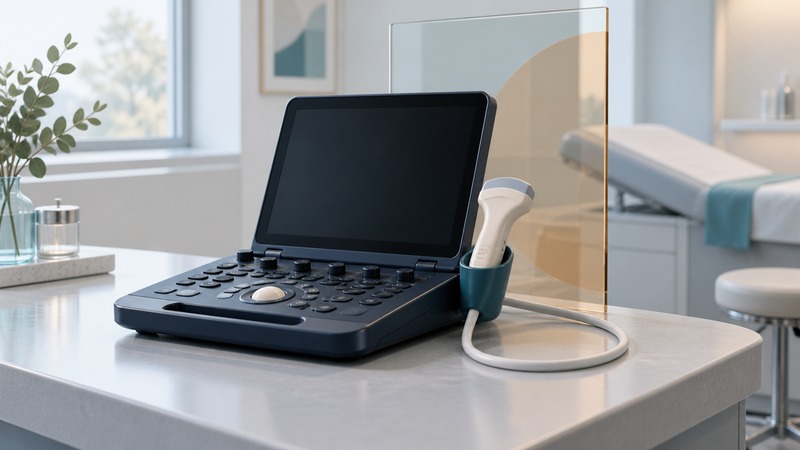
A small machine may support a focused bedside assessment or a more extensive examination. Its size alone tells you little about the scope of the test. The clinical question, the operator's training, the probe, and the examination protocol all matter.
Match the scan to the question
RadiologyInfo, from the ACR and RSNA, explains general ultrasound and the use of Doppler to evaluate blood flow. An echocardiogram is an ultrasound examination of the heart. A scan used to guide a procedure has a different purpose from a diagnostic survey of an organ.
On a small screen, scroll the table sideways to read all columns.
| Term | What it tells you | Question to ask |
|---|---|---|
| Diagnostic ultrasound | Images of a specified area to investigate a clinical question. | Which organ or problem is being assessed? |
| Doppler | Additional information about blood movement. | Is blood-flow assessment part of my ordered examination? |
| Point-of-care ultrasound | An assessment performed where care is being given, often answering a focused question. | What does this scan establish, and is a comprehensive study also needed? |
| Ultrasound guidance | Images help a clinician guide a separate procedure. | What preparation, consent and aftercare does that procedure require? |
The American Institute of Ultrasound in Medicine describes bedside applications. Its point-of-care practice parameter also describes circumstances requiring comprehensive imaging. A focused answer should be interpreted within the examination actually performed.
Confirm preparation with the imaging unit
Some examinations require fasting; others call for a full bladder. Follow the instructions for your booked scan. RadiologyInfo explains why preparation differs. Confirm the start time for any restriction, what you may drink, and what to do about medicines. Tell the unit if fasting or drinking extra fluid conflicts with an existing diabetes, kidney or other care plan.
Have the referral and appointment details ready when calling. Say exactly which instruction is unclear. Ask about accessibility, help with positioning, an interpreter, or support for a child before the visit. If you have already eaten or could not complete the preparation, tell the unit so it can decide how to proceed.
What happens in the room
For an external scan, the operator usually applies gel and moves a handheld probe over the area. You may be asked to change position or briefly hold your breath. Pressure over a tender area can be uncomfortable; tell the operator so they can adjust the examination. Some scans use an internal probe: confirm whether this is planned and discuss consent, comfort and a chaperone.
Leave with a follow-up route
Ultrasound uses no ionizing radiation. Its usefulness still depends on what can be seen: air, bone and the depth of the target can limit images. An incomplete view may lead to additional imaging. Ask what was adequately assessed and what remains uncertain.
A radiology examination generally produces a report for the requesting clinician. Confirm who will explain it, when to expect contact, and whom to call if the agreed time passes. A quiet examination room or a saved screen image is not a result. Use the medical-records guide if another clinician needs both the report and original study.
Download the ultrasound appointment planner to record the exact instructions and follow-up arrangement. For a different imaging appointment, see the MRI preparation guide.
General patient education. Use your clinical team’s instructions for your care. Explore the medical guides.