The strongest brain-mapping workflows in 2026 are not one-model tricks. They are layered pipelines that combine segmentation, registration, denoising, atlas alignment, quality control, multimodal fusion, graph analysis, and expert review across MRI, fMRI, diffusion imaging, electron microscopy, and molecular atlases. That is what makes modern connectome and atlas work feel materially different from older neuroimaging automation.
The current ground truth is unusually strong. Nature published BrainParc on March 11, 2026 as a unified lifespan parcellation model from structural MRI, Nature Computational Science published deepmriprep in February 2026, Nature published the NextBrain probabilistic histological atlas in December 2025, MICrONS published large-scale structure-function mapping of mouse visual cortex in April 2025, and the adult fruit fly whole-brain wiring diagram landed in Nature in October 2024. These are not generic AI demos. They are core infrastructure papers.
That also means this page has to stay careful. Some advances here are clearly operational in research pipelines today, while others remain translational or exploratory for clinics. A strong 2026 page on brain mapping should say where AI is already reliable, where multimodal learning is expanding the map, and where human-in-the-loop review still matters.
1. Automated Image Segmentation
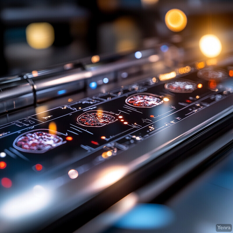
Automated segmentation is now core infrastructure for brain mapping, not an optional convenience. The practical change is that whole-brain delineation, tissue labeling, and region boundary extraction can now happen quickly enough to support large MRI and histology programs without collapsing under manual annotation cost.
OpenMAP-T1 is a strong operational anchor because it reports rapid deep-learning parcellation of 280 brain regions, while NextBrain shows how multimodal ex vivo MRI and histology can support probabilistic whole-brain labeling at higher fidelity. Inference: segmentation is strongest in 2026 when it is tied to atlas quality, robustness, and downstream reproducibility rather than only dice scores.
2. High-Resolution Connectome Reconstruction
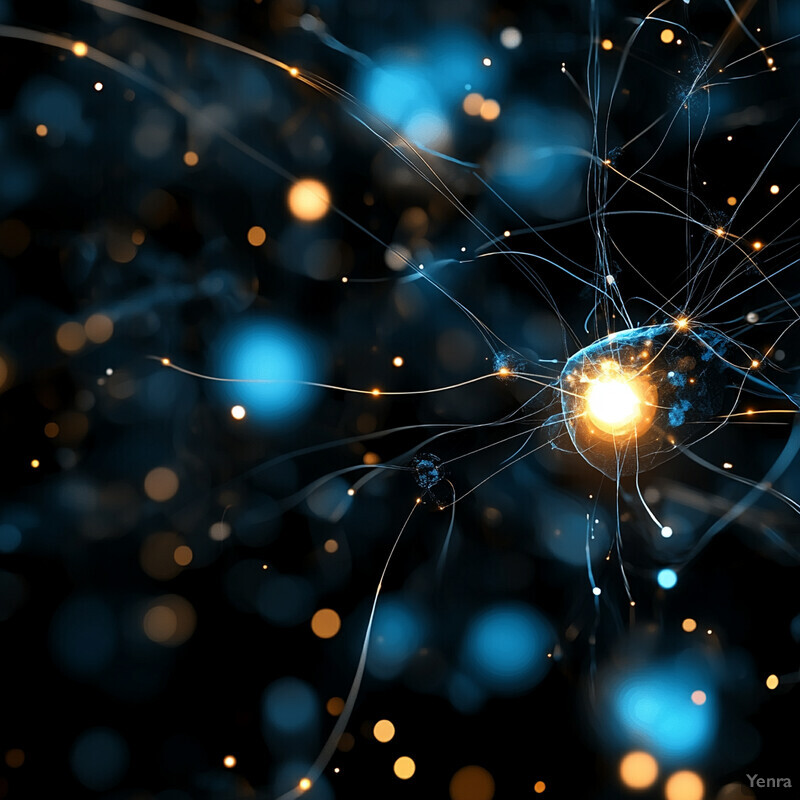
High-resolution connectome reconstruction has moved from partial proof-of-concept to whole-system mapping in selected organisms and cortical volumes. AI is central because tracing neurites and synapses across petascale image stacks is still too large and too error-prone for manual reconstruction alone.
The adult fruit fly whole-brain wiring diagram and the 2025 MICrONS structure-function paper are the clearest grounding points here. Together they show that AI-enabled reconstruction is now good enough to support dense synapse-level maps and then relate them to function. Inference: the modern bottleneck is no longer whether tracing can be automated at all, but how reliably it can be proofread, aligned, and interpreted.
3. Accelerated Image Processing
Acceleration is one of the most concrete AI wins in neuroimaging. Faster preprocessing matters because large-scale imaging cohorts are no longer useful if surface reconstruction, alignment, and normalization remain slow enough to delay the science by months.

DeepPrep showed large-scale acceleration in 2025, and deepmriprep extended that argument in 2026 by packaging a faster deep-learning-first preprocessing workflow with broad dataset support. Inference: acceleration is no longer just about speed for its own sake; it is what makes repeated QC, atlas comparison, and cohort-scale reanalysis practical.
4. Improved Noise Reduction
AI denoising is getting stronger because it can model artifacts more flexibly than fixed filters, especially in EEG and other noisy physiological recordings. The useful boundary is that good denoising should preserve neural structure instead of polishing away the signal of interest.

AnEEG is a strong anchor because it focuses on EEG denoising without paired clean labels, and a 2025 Scientific Reports model pushed artifact removal further with transformer-style attention. Inference: denoising is strongest when teams evaluate what the model preserved, not just how smooth the output looks.
5. Multi-Modal Integration
Multi-modal integration is where brain mapping becomes far more informative than any single scan type. Structure, function, diffusion, transcriptomics, and molecular atlases answer different questions, and AI helps put them into the same analytical frame instead of leaving them as parallel silos.

The Human Connectome Project remains a foundational official anchor for multimodal mapping, while GIANT shows how graph-based integration can connect imaging, phenotype, and genetic information. Inference: multimodal brain mapping is most valuable when it preserves the differences between modalities while still learning the correspondences among them.
6. Automated Brain Parcellation
Parcellation is improving because AI can now learn region boundaries directly from population and subject-specific data rather than only inheriting a fixed atlas. That matters for research quality and for any workflow that needs individualized rather than averaged brain maps.
BrainParc is a major 2026 anchor because it frames parcellation as a unified lifespan task from structural MRI, while recent HCP-based individual cortical parcellation work shows subject-level mapping is becoming more practical at scale. Inference: the field is moving from “which atlas?” toward “which atlas plus which individual adjustment?”
7. Predictive Modeling of Brain Functions
Predictive modeling is where static brain maps start to become mechanistic tools. The important shift is not just classifying conditions from scans, but using mapped activity and connectivity to forecast how circuits behave during tasks and decisions.
The 2025 International Brain Laboratory paper is a strong anchor because it maps brain-wide activity during complex behavior rather than a narrow toy task, while MICrONS helps link that predictive ambition to dense structure-function correspondence. Inference: prediction gets more credible when models are grounded in richer maps rather than asked to infer function from sparse summary features.
8. Advanced Feature Extraction
Advanced feature extraction is moving from hand-crafted imaging summaries toward learned representations that can travel across studies. That matters because brain-mapping pipelines increasingly need features that remain useful across scanners, cohorts, and downstream tasks rather than only one narrow benchmark.

Brain Graph Foundation Model and BrainSymphony are good 2025 anchors because both push toward transferable brain representations instead of one-task classifiers. Inference: feature extraction is strongest in 2026 when it is treated as shared infrastructure for many neuroimaging tasks, not just a trick to boost one leaderboard.
9. Early Biomarker Detection
Brain-mapping AI is contributing to earlier biomarker work, but the strongest claims are still about risk stratification and enriched signal detection rather than fully autonomous diagnosis. Good biomarker models usually combine mapping quality, multimodal context, and longitudinal validation.

Recent Nature Communications work on multimodal human brain age and longitudinal brain aging gives this section stronger footing because it ties biomarker detection to interpretable deviation from expected trajectories. Inference: the strongest near-term value of these models is early warning and cohort stratification, especially for neurodegeneration research.
10. Adaptive Registration and Alignment
Registration and alignment remain foundational because every downstream map depends on whether signals from different brains, modalities, or histological slices actually line up. AI helps most when it improves robustness to distortion, damage, or modality mismatch rather than just making standard cases slightly faster.

NextBrain is a particularly good anchor here because its probabilistic atlas depends on careful multimodal alignment of ex vivo MRI and histology, while deepmriprep shows how registration quality now sits inside faster, more automated pipelines. Inference: alignment is still one of the hidden determinants of whether a brain map becomes scientifically reusable.
11. Quantitative Analysis of Microcircuitry
AI is making microcircuit analysis more quantitative by turning dense microscopy and cell-atlas data into counts, motifs, and cell-type-resolved circuit descriptions. That is a major step up from simply viewing beautiful imagery without being able to measure it consistently.

MICrONS gives this section strong structure-function grounding, while the 2025 whole-mouse-brain cellular atlas shows how AI-assisted mapping can quantify cell classes and their spatial organization across the entire brain. Inference: modern brain mapping is increasingly about statistics on microcircuitry, not only image reconstruction.
12. Automated Quality Control
Quality control has become more important, not less, as pipelines automate. Fast mapping only helps if teams can detect motion, scanner artifacts, failed surfaces, mislabeled parcels, and subtle bias before those errors contaminate downstream statistics.
The 2025 Nature Neuroscience paper on artifactual bias in automated MRI analyses is an important cautionary anchor, and recent JMRI work on rank-based ratings shows how QC can be formalized rather than left to ad hoc inspection. Inference: a stronger brain-mapping pipeline in 2026 is usually a more self-critical one.
13. Human-in-the-Loop Systems
Human oversight still matters because the hardest failures in brain mapping are often rare, local, and scientifically expensive. In connectomics and closed-loop neurophysiology especially, AI is strongest when it accelerates experts instead of pretending proofreaders, anatomists, and experimentalists are obsolete.

Autoproof is a current connectomics anchor because it focuses directly on proofreading assistance, while NeuroART shows how real-time neurophysiology systems still depend on researchers staying inside the analytical loop. Inference: human-in-the-loop design is not a fallback here; it is part of normal scientific operations.
14. Graph Analytics for Connectomics
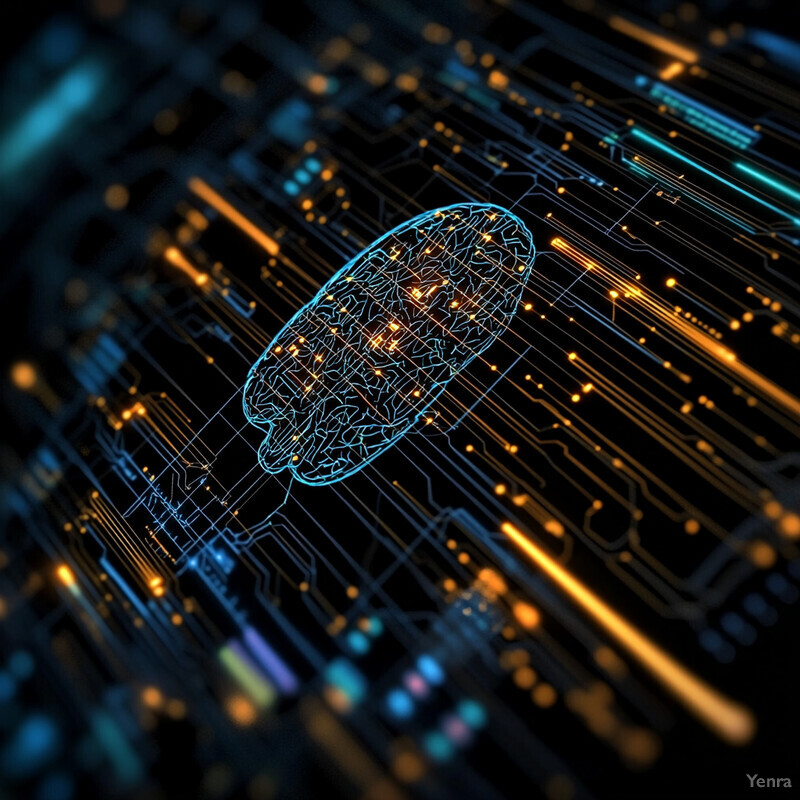
Graph analytics matters because brain maps are relational objects. Region-to-region connectivity, synaptic wiring, and cell-type interaction all benefit from graph-aware models rather than workflows that flatten networks into disconnected features.

The 2025 npj AI perspective is useful because it reframes functional connectome analysis around graph deep learning rather than older summary statistics alone, and Brain Graph Foundation Model provides a more direct current modeling example. Inference: graph methods are becoming central because the brain is genuinely graph-structured, not because GNNs are fashionable.
15. Reduction of Manual Labeling Effort
Label-efficient learning matters because expert annotation remains one of the most expensive parts of brain mapping. The strongest progress here comes from active learning, semi-supervised training, and representation learning that let scarce human labels stretch much further.

SAND is a strong anchor because it explicitly combines semi-supervised learning with active selection of useful annotations in calcium imaging, while CellTransformer shows how self-supervised spatial modeling can reduce dependence on hand-crafted region labels in atlas-building work. Inference: the field is steadily replacing brute-force labeling with smarter supervision.
16. Causal Inference in Connectivity
Causal inference is still one of the more exploratory parts of AI brain mapping, but it matters because researchers want more than correlation maps. They want models that can say something defensible about directional influence, perturbation effects, and mechanistic circuit hypotheses.
A 2025 dynamic causal discovery preprint is a good current anchor because it tackles directionality in time-varying neural data directly, while recent TMS-fMRI work shows how causal connectome ideas can be tested against perturbation data rather than only resting correlations. Inference: the promising direction here is combining AI discovery with intervention-rich datasets, not claiming causality from observation alone.
17. Integration with Genomic Data
Imaging-genomics integration is getting stronger because molecular atlases are becoming detailed enough to line up with mapped brain structure and connectivity. AI helps bridge those scales, from gene expression and cell type to region-level imaging phenotypes.

GIANT is a strong 2025 anchor because it explicitly builds a genetically informed atlas, while the 2024 Brain Cell Atlas shows how cell-type-resolved, whole-brain molecular mapping is becoming a serious reference layer for imaging interpretation. Inference: the next decade of brain mapping will be much more molecularly grounded than the last one.
18. Personalized Brain Mapping
Personalized brain mapping is becoming more practical because models can now adapt population atlases to the individual instead of forcing every brain through one template. That is especially important for longitudinal monitoring, surgical planning, and precision-neuroscience research.
The HCP individual cortical parcellation paper is a direct anchor because it targets subject-level mapping, and the 2026 Nature Communications brain-aging work strengthens the longitudinal side by improving individual trajectory estimates. Inference: personalized mapping is strongest when it combines individualized structure with repeated measures over time rather than relying on one scan alone.
19. Virtual and Augmented Reality for Visualization
Visualization still matters because large brain maps are hard to interpret on flat screens. The practical value of immersive tools is not novelty. It is making dense network structure, tract geometry, and multi-layer atlas relationships easier for researchers and clinicians to inspect together.
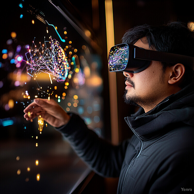
NeuroCave remains a useful research anchor because it shows immersive connectome exploration as a serious analytical interface, while Connectome Workbench remains an operational visualization tool in large consortium workflows. Inference: the strongest visualization systems are the ones that keep linked metrics, atlases, and interaction grounded in the underlying data.
Sources and 2026 References
- BrainParc grounds current lifespan parcellation from structural MRI.
- deepmriprep and DeepPrep ground the current preprocessing and acceleration stack.
- NextBrain grounds multimodal histological atlas building and high-fidelity alignment.
- OpenMAP-T1 grounds fast large-scale whole-brain segmentation and parcellation.
- the adult fruit fly brain wiring map and MICrONS structure-function mapping ground current connectomics scale.
- A brain-wide map of neural activity during complex behaviour grounds current whole-brain functional mapping at behavioral scale.
- AnEEG and recent EEG artifact-removal work ground AI denoising.
- the Human Connectome Project and the HCP-YA 2025 release materials ground current multimodal mapping infrastructure.
- Brain Graph Foundation Model and BrainSymphony ground current representation-learning and feature-extraction trends.
- Genetic architecture of multimodal human brain age and Precision estimates of longitudinal brain aging ground early biomarker and individualized aging analyses.
- Artifactual bias in automated MRI analyses and rank-based MRI quality ratings ground the current QC and bias conversation.
- Autoproof and NeuroART ground human-in-the-loop and closed-loop research workflows.
- Rethinking functional brain connectome analysis with graph deep learning grounds current graph-analytics framing.
- SAND and CellTransformer ground label-efficient learning and atlas construction.
- dynamic causal discovery from neural time series and TMS-fMRI causal connectome work ground the current causal-inference boundary.
- GIANT, Brain Cell Atlas, and the molecularly defined cellular atlas of the entire mouse brain ground imaging-genomics and cell-atlas integration.
Related Yenra Articles
- Brain-Computer Interfaces (BCI) shows how mapped neural signals become interactive systems.
- Biomarker Discovery in Healthcare connects brain-mapping outputs to translational marker development.
- Patient Outcome Prediction extends mapped brain features into prognosis and longitudinal risk models.
- Personalized Medicine provides the clinical companion for individualized mapping and treatment planning.