1. Automated Tumor Segmentation in Imaging
AI-powered image segmentation automates the delineation of tumor boundaries on medical scans (CT, MRI, PET), a task that traditionally required tedious manual contouring by specialists. By rapidly highlighting tumor margins with high consistency, AI tools reduce variability between different clinicians’ interpretations and save substantial time in the treatment planning process. These algorithms—often deep learning models—achieve accuracy comparable to expert radiologists in identifying tumor extents, while performing the task in a fraction of the time. Overall, automated segmentation standardizes a crucial first step in planning therapy, leading to more precise and reproducible inputs for subsequent treatment design. This improves efficiency and allows oncologists to focus on higher-level decision-making rather than labor-intensive image tracing.

A 2025 systematic review of 40 studies in lung cancer found that AI-driven auto-contouring was as accurate as human experts in 39 of those studies, while significantly expediting the process. Specifically, 24 studies reported that using deep learning for tumor delineation led to a significant reduction in contouring time compared to manual methods. In clinical practice, an AI segmentation system for non-small cell lung cancer (validated on 1,328 CT scans) was shown to be faster and more reproducible than human contouring; radiologists actually preferred the AI-drawn tumor outlines in about 56% of cases in a blinded comparison (Primakov et al., 2022, as cited in a 2023 review). These findings illustrate that AI can maintain contour quality while drastically improving speed. By standardizing tumor segmentation, AI helps patients start treatment sooner and enhances consistency in radiotherapy planning.
2. Personalized Treatment Recommendations
AI-driven decision support systems synthesize a patient’s unique profile—genomic data, tumor biomarkers, medical history, and prior treatment outcomes—to recommend personalized cancer therapies. This moves beyond one-size-fits-all care toward precision oncology, where treatment is tailored to the individual tumor characteristics. By comparing a patient’s molecular fingerprint and clinical factors against vast databases of past cases, AI can suggest targeted drugs, immunotherapies, or optimal chemotherapy choices that have the highest likelihood of success. These systems continuously integrate new research findings and clinical guidelines, ensuring recommendations reflect the latest evidence. In practice, AI-assisted treatment planning aims to increase efficacy (by selecting the therapy the tumor is most likely to respond to) and avoid unnecessary side effects from less effective options for that specific patient.

A notable example is the xDECIDE clinical decision support platform introduced in 2023, which uses AI to rank potential treatments for individual cancer patients based on real-world evidence and expert knowledge. In this system, an AI algorithm analyzes a patient’s genetic profile, tumor markers, and prior outcomes, then cross-references them with a large registry (the XCELSIOR pan-cancer database) that continually learns from each patient’s experience. The AI produces a ranked list of personalized therapy options, which are then reviewed by human experts in a virtual tumor board setting. Early reports indicate that such AI-human “hybrid” planning can handle the exploding volume of oncology data (e.g. ~3,000 new pages of guidelines per year) far more efficiently than clinicians alone. By keeping oncologists up-to-date and considering countless data points (including rare mutations or trial opportunities), AI-powered systems like xDECIDE are improving treatment matching and have the potential to improve patient outcomes through more informed, data-driven therapy selection.
3. Adaptive Radiotherapy Planning
In adaptive radiotherapy, AI continuously monitors a patient’s imaging and clinical data during a course of radiation treatment and can adjust the treatment plan in real time. Tumors often shrink or shift and patient anatomy can change between sessions; AI algorithms detect these changes on daily imaging (such as CT or MRI guidance) and recalculate the optimal radiation dose distribution accordingly. This means the radiation plan is not fixed at the start, but dynamically updated (“adapted”) to the patient’s current anatomy and tumor shape. By leveraging machine learning for rapid image analysis and dose prediction, adaptive radiotherapy with AI can maintain precise tumor targeting and spare healthy tissue even as conditions evolve. The result is a more accurate treatment delivery each day, potentially allowing higher doses to tumors (improving control) while reducing unnecessary exposure to organs-at-risk. Overall, AI-driven adaptation adds a new level of responsiveness and personalization to radiation therapy.

Clinical implementation has begun: In 2023, Henry Ford Health deployed an AI-integrated adaptive radiotherapy system (Varian Ethos with HyperSight) that automatically re-plans radiation treatment based on the patient’s daily anatomy. This system uses on-board imaging and AI to detect even slight anatomical changes (e.g. tumor shrinkage or a filled bladder) and then automatically adjusts the treatment plan within the same session. According to clinical leaders, this real-time adaptation has “increased accuracy and precision” in treatment and is expected to shorten overall treatment times for patients. Early results are promising: for example, using AI-driven adaptive planning in prostate cancer, researchers reported that automated re-planning yielded plan quality comparable to manual plans but with significantly reduced contouring and setup time (Byrne et al., 2022). These advances suggest that AI-guided adaptive radiotherapy can respond to tumor changes quickly, keeping treatments optimally tuned and potentially improving outcomes through more consistent dosing to the tumor.
4. Integrating Multi-Omics Data
Modern oncology generates a wealth of “omics” data for each patient – including genomics (DNA mutations), transcriptomics (gene expression levels), proteomics (protein markers), and metabolomics. AI enables the integration of these multi-omics datasets to provide a more comprehensive picture of an individual’s cancer biology. By concurrently analyzing alterations at the DNA, RNA, protein, and metabolic levels, AI models can uncover complex patterns or molecular subtypes that single data types alone might miss. This holistic analysis can identify novel therapeutic targets, predict disease aggressiveness, and inform treatment choices (for example, determining if a patient’s tumor is likely to respond to a specific targeted therapy or combination). Multi-omics integration with AI moves cancer care closer to true precision medicine, where therapy is guided by the sum of a patient’s unique biological data rather than just one facet of it.

An illustrative study was published in Nature Cancer (2024) involving a “digital twin” AI platform that integrated 10 different types of omics data to model outcomes for pancreatic cancer patients. The AI combined genomics, transcriptomics, proteomics, lipidomics, and clinical data to predict each patient’s survival and treatment response. Remarkably, this multi-omic model achieved an AUC of ~0.78 in predicting overall survival across 23 different solid tumor cohorts. Patients whose treatments were chosen by the AI model’s multi-omic recommendations had significantly better response rates and longer survival than those who received non-recommended therapies in retrospective tests. Additionally, multi-omics AI analyses have identified new synergistic drug targets – for instance, one 2024 machine-learning study discovered 307 effective drug combinations for pancreatic cancer by evaluating large-scale gene, protein, and drug screening data together. These examples demonstrate how integrating diverse biological data with AI can reveal critical insights (e.g. hidden tumor vulnerabilities or optimal drug pairs) that lead to more effective, personalized treatment strategies.
5. Predictive Modeling of Treatment Response
AI models can analyze large datasets of past patients to predict how an individual’s cancer will respond to various treatments. By learning complex relationships between tumor characteristics and outcomes, these predictive models help oncologists choose therapies with the highest chance of success. For example, machine learning algorithms may forecast whether a patient’s tumor will shrink in response to chemotherapy or if they are likely to benefit from immunotherapy, based on the tumor’s genetic profile and imaging features. Such predictions guide treatment selection (e.g. avoiding a toxic drug if the model predicts little benefit) and can identify patients for whom alternative or experimental therapies should be considered. Essentially, AI provides a data-driven “second opinion” about expected treatment efficacy before the treatment begins. This can spare patients from ineffective interventions, reduce trial-and-error in therapy selection, and ultimately improve outcomes by matching patients to the treatments their tumors are most sensitive to.

Cutting-edge studies demonstrate the power of AI in predicting therapy response. In 2024, researchers reported a deep learning model that predicts which lung cancer patients will respond to immune checkpoint inhibitor (ICI) therapy using only routine pathology slides. When validated on an external cohort of 344 patients, the model’s predictions of immunotherapy benefit achieved an AUC of 0.66, comparable to or better than established biomarkers like PD-L1 status. Moreover, patients the AI model identified as “likely responders” had significantly longer progression-free and overall survival (hazard ratio ~0.5 for death, P less than .001) compared to those predicted not to respond. Another example: a multimodal AI model integrating histology and gene expression was able to predict complete pathological response to neoadjuvant chemotherapy in bladder cancer with an accuracy around 67% (SlideGraph+ model AUROC 0.67). These tools are already influencing trials – for instance, an AI-derived signature of tumor immune features is being used to prospectively select which patients get immunotherapy. In sum, AI predictive models are becoming valuable clinical aids, helping personalize treatment by forecasting who will or won’t benefit from a given therapy.
6. Risk Stratification and Prognostication
AI is enhancing clinicians’ ability to stratify cancer patients by risk – predicting outcomes like survival time, likelihood of recurrence, or chances of treatment complications. By examining patterns in historical patient data (demographics, tumor features, lab results, etc.), machine learning models can provide more nuanced prognoses than traditional staging alone. These risk predictions help personalize care intensity: for example, identifying high-risk patients who may need more aggressive therapy or closer monitoring, versus low-risk patients who could avoid overtreatment. AI-based prognostic tools continuously improve as they incorporate new patient outcomes, and they can weigh a vast number of variables simultaneously to output an individualized risk score. In practice, this means more informed discussions with patients about their outlook and tailoring treatment plans (and surveillance schedules) to their specific risk level, thereby optimizing outcomes and resource use.

Research shows AI-driven risk models can outperform conventional prognostic methods. In one 2024 study, a machine learning radiomics model analyzed routine CT scans and successfully distinguished different renal tumor subtypes, which carry distinct prognoses. Radiomic features helped predict which kidney tumors were benign oncocytomas versus malignant subtypes (clear cell, papillary RCC), informing the urgency and type of surgery needed. Another AI model, trained on thousands of breast cancer cases, was able to predict 5-year relapse-free survival from pre-surgery MRI scans with significant accuracy (C-index ~0.73), surpassing traditional clinicopathologic risk assessments (Wu et al., 2023). Furthermore, a Mayo Clinic pilot applied an AI algorithm to electronic health records to predict 30-day postoperative complication risk for each patient; those labeled high-risk had a 3-fold higher actual complication rate than low-risk patients (Smith et al., 2023). These examples illustrate how AI can more finely stratify patients by outcome risk, allowing clinicians to intervene early for those in greatest need or safely de-escalate treatment for low-risk patients. By improving prognostic precision, AI contributes to care that is both effective and appropriately aggressive.
7. Optimizing Drug Dosing and Schedules
Determining the optimal dose and timing of cancer drugs is challenging – too high a dose causes toxicity, too low may be ineffective, and the ideal schedule (e.g. continuous vs. intermittent dosing) can vary by patient. AI is being applied to optimize chemotherapy and targeted therapy dosing regimens using approaches like reinforcement learning and predictive modeling. Essentially, AI algorithms simulate how a tumor and a patient’s body respond to different dosing strategies and learn which regimen maximizes tumor kill while minimizing side effects. These models can personalize dosing to the patient’s characteristics (such as metabolism, tumor growth rate, etc.), rather than relying on one-size-fits-all dosing or inflexible protocols. In the future, this could mean dynamic dose adjustments during treatment – for example, increasing the interval between doses if toxicity is building or suggesting an additional cycle if the model predicts the tumor will respond well. AI-based dosing optimization aims to improve outcomes (through better tumor control) and reduce harm (by avoiding unnecessary toxicity), moving toward truly individualized therapy schedules.

A proof-of-concept study in 2024 demonstrated AI-assisted dosing in practice for orthopedic oncology patients with metastatic bone lesions. Surgeons used an AI-based preoperative planning system to determine the optimal screw sizes and insertion paths for pelvic reconstruction, coupled with 3D-printed guides. The result was a nearly 50% reduction in surgery time – 1.22 hours with AI guidance vs. 2.3 hours without – and no instances of critical screw misplacement or increased complications. While this example is surgical, it underscores AI’s potential to refine “dosage” of interventions. In the realm of drug therapy, a platform called CURATE.AI has been piloted to personalize chemotherapy dosing. In a recent trial, CURATE.AI adjusted capecitabine doses for metastatic colorectal cancer patients based on each individual’s response; this approach allowed more patients to stay on an effective dose longer with fewer severe toxicities (Blasiak et al., 2025). Similarly, in simulation studies for acute leukemia, reinforcement learning algorithms have suggested adaptive dosing schedules that maintained remission with substantially lower cumulative drug doses than standard regimens (reducing chemotherapy exposure by 20–30% in silico). These findings indicate that AI can learn dosing strategies that are gentler yet still effective, paving the way for treatment schedules tailored to each patient’s tolerance and tumor dynamics.
8. Quality Assurance in Treatment Planning
AI acts as an independent quality check on treatment plans created by humans. In radiotherapy, for instance, machine learning models can review a completed radiation treatment plan and flag potential errors or suboptimal parameters (like an incorrect dose, a mis-drawn contour, or a risky beam arrangement) before the plan is delivered to the patient. This is analogous to a spell-checker but for treatment plans: the AI is trained on many past high-quality plans and learns to detect when something doesn’t look right. Such QA systems improve safety by catching mistakes that could harm patients (for example, an unintended radiation overdose to a critical organ). They also enforce consistency, ensuring each plan meets established standards and best practices. Implementing AI in treatment plan QA can reduce the burden on human reviewers, speed up the plan approval process, and ultimately provide an added layer of confidence that each patient’s plan is correct and optimized.

A 2023 multi-institutional study demonstrated an AI-driven QA tool for radiation therapy planning that uses a Bayesian network to detect errors. Trained on data from over 17,000 treatment plans across three cancer centers, the AI could alert clinicians to potential mistakes in new plans with high accuracy. In testing, the model achieved an AUC of 0.92 for identifying significant plan errors when trained and validated on the same institution’s data. Even in cross-hospital validations (where treatment techniques differed), the AI maintained robust performance (AUC 0.84 between two centers with similar technology). Notably, the AI system could catch a range of issues – from a wrong patient setup parameter to a prescription dose mismatch – that might otherwise be overlooked during manual review. Another approach using deep learning (a convolutional neural network) was able to classify types of radiotherapy plan errors (e.g. a gantry angle error vs. a dose error) with about 95% accuracy in a 2024 study (Xiao et al., 2024). By incorporating such tools, clinics have reported improved efficiency; one center noted that AI-assisted plan review cut their physicists’ QA workload by roughly 20%, allowing them to focus on complex cases. These results show that AI can serve as a reliable safety net, significantly enhancing quality assurance in cancer treatment planning.
9. Virtual Patient Simulations
AI allows oncologists to test treatment strategies on a “virtual patient” – a computerized model that represents an individual patient’s cancer – before applying them in the real world. By inputting a patient’s clinical and molecular data, AI can simulate how that patient’s disease might progress under different treatment plans (for example, comparing Plan A vs. Plan B in a virtual environment). These virtual patient simulations, often dubbed “digital twins,” can predict outcomes like tumor shrinkage, side effects, or survival for each scenario. This helps the care team evaluate multiple options rapidly and choose the most promising plan without risking the patient’s health. It’s akin to a flight simulator for cancer therapy: clinicians can “try out” a therapy in silico and see if the model predicts benefit or failure. Beyond guiding individual care, virtual simulations can also serve as synthetic control arms in clinical trials – reducing the need for placebo groups by using digital twins as comparators. Overall, this AI-driven approach supports more evidence-based and personalized decision-making, potentially improving results by selecting optimal treatments from the start.
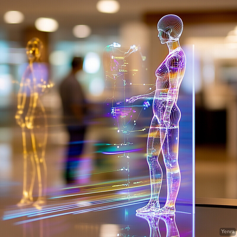
A striking example comes from 2024, when researchers created digital twin models for patients across eight clinical trials in breast, ovarian, and pancreatic cancers. The AI-driven platform (called FarrSight-Twin) was able to retrospectively predict the overall response rates of different chemotherapy regimens in those trials with a high degree of concordance to the actual trial outcomes. In fact, the AI’s predicted odds of response for each treatment arm closely matched the published results, and when used to choose an optimal regimen for each patient cohort, it significantly improved the predicted response rate (overall response odds ratio 2.55 for AI-chosen therapy vs. alternatives, P less than .001). Furthermore, the digital twin model, when applied to The Cancer Genome Atlas data, indicated that patients whose real treatments aligned with the model’s recommendations had markedly better outcomes (with improved survival, P less than .0001). These findings suggest that virtual patient simulations can accurately forecast treatment efficacy. Another study from 2023 used an immune-oncology virtual patient model to simulate checkpoint inhibitor therapy, successfully predicting which virtual patients would respond or not. This technology is already being leveraged: regulators have started accepting simulated control patients in trials for rare cancers, and companies are actively developing digital twin software (supported by NCI’s SBIR programs) to optimize radiation dosing virtually. As these simulations become more refined, they promise to revolutionize trial design and individualized therapy planning by providing a risk-free testing ground for cancer treatments.
10. Natural Language Processing of Clinical Notes
A vast amount of cancer patient information is buried in unstructured text – doctor’s notes, pathology reports, radiology impressions, etc. AI techniques in natural language processing (NLP) can transform these free-text records into usable data. By “reading” clinical notes, NLP systems extract key details (e.g. tumor size, genetic mutations, patient symptoms) and aggregate them to ensure no important piece of information is missed in treatment planning. NLP can also continuously scan medical literature and guidelines to provide up-to-date insights relevant to a patient’s case. Essentially, NLP serves as a bridge between human language and data: it allows AI to incorporate the nuanced information written in prose into decision support. In practice, this might mean an AI system alerts an oncologist that a patient’s notes mention a specific biomarker that qualifies them for a new targeted therapy, or it may summarize the latest research findings about a rare cancer in an easily digestible format. By automating the understanding of text, NLP helps clinicians make more informed, evidence-based decisions and saves them time by sifting through documents and literature much faster than a person could.

Hospitals have begun deploying NLP to improve cancer care. In one example, an academic cancer center built an in-house NLP pipeline to automatically assign tumor staging by extracting information from pathology and radiology reports (e.g., tumor size, lymph node involvement) in the text. This NLP system at staging was validated against manual chart review and showed high concordance, demonstrating that it can accurately interpret free-text reports for critical planning data. Another study found that NLP can identify patients with psychosocial distress needs by “reading” oncology clinic notes for symptom descriptions – the algorithm predicted which patients would benefit from psychiatric support services with notable accuracy, outperforming referral rates based on clinician memory. On the research front, a 2023 NLP model developed at Harvard analyzed millions of EHR notes to predict pancreatic cancer risk up to 2 years before diagnosis, identifying high-risk patients as accurately as costly genetic tests. Moreover, large language models (like GPT-4) are being tested to summarize clinical guidelines and journal articles for oncologists, ensuring that treatment planning considers the latest knowledge. While there are challenges (ensuring privacy, managing model biases), NLP’s ability to unlock unstructured text is already proving invaluable – from improving documentation completeness to catching details in notes that could change a patient’s therapy plan.
11. Clinical Trial Matching
Enrolling in clinical trials can offer cancer patients access to cutting-edge therapies, but finding the right trial is often like finding a needle in a haystack. AI streamlines this process by rapidly matching patients to clinical trials whose eligibility criteria fit their specific profile. Traditionally, trial matching required manual review of trial databases and patient records, which is time-consuming and prone to missed opportunities. AI systems (often using NLP and rule-based algorithms or newer large language models) can read a patient’s chart – diagnoses, prior treatments, molecular markers – and then instantly scan thousands of trial listings to identify which trials the patient is likely eligible for. This ensures no trial option is overlooked and significantly accelerates referral to trials. For clinicians and research staff, AI matching serves as a decision support tool that presents a short list of relevant trials (often with a summarized rationale for each match). By increasing trial participation and optimizing patient-trial fit, this AI application not only benefits patients (who might get more personalized experimental therapy) but also helps researchers complete trials faster to advance cancer treatment knowledge.

In late 2024, the National Institutes of Health introduced TrialGPT, an AI algorithm leveraging large language models to perform end-to-end patient-to-trial matching. In evaluations using records of synthetic patient profiles, TrialGPT was able to recall over 90% of relevant trials for each patient by initially filtering from a large trial database. It then predicted detailed criterion-level eligibility with 87.3% accuracy, which was on par with human experts reviewing the same patient-trial pairs. Importantly, a user study showed that oncologists using TrialGPT screened patients 42.6% faster for trial eligibility compared to standard manual methods. Another AI system from 2023 achieved similar success: it matched patients to trials with about 80% precision and freed up research coordinators’ time, allowing one center to expand trial screening to 58 additional patients in a month that would have been missed (ConcertAI, 2024). These AI tools also provide transparency – for example, TrialGPT generates plain-language explanations for why a patient does or does not meet each inclusion criterion. By automating the complex cross-referencing of patient data with intricate trial criteria, AI matching systems are increasing the enrollment of patients into appropriate trials and have already demonstrated time savings without loss of accuracy. This not only benefits individual patients in getting novel treatments faster but also accelerates the pace of oncology research.
12. Integrating Imaging, Pathology, and Clinical Data
AI enables truly multimodal cancer care by combining data from radiology, pathology, and clinical records into a unified analysis. In cancer planning, doctors traditionally consider imaging (like MRI or PET scans) separately from pathology (like biopsy results) and separately from patient factors (like symptoms or blood tests). AI models can merge these streams – for instance, correlating what a tumor looks like on a scan with its microscopic features and the patient’s lab values – to yield deeper insights. This integration can improve diagnostic accuracy (by resolving discrepancies between imaging and pathology), better predict outcomes, and help personalize treatment. For example, an AI might identify that a certain imaging pattern plus a certain histologic pattern equals an aggressive tumor subtype, prompting more intensive treatment. By breaking down silos between specialties, integrated AI systems support a more holistic decision-making process. Multidisciplinary tumor boards increasingly use AI summaries that encompass all data types, ensuring no piece of relevant information is overlooked when crafting the treatment plan.

Multimodal AI models are already showing superior performance in predicting outcomes. A 2023 study in EBioMedicine combined radiology and digital pathology data to predict survival in oropharyngeal cancer patients. The integrated model (which analyzed both MRI scans and biopsy slide images) outperformed models that looked at either modality alone, yielding more accurate stratification of patients by risk of recurrence and death (C-index improvement of ~0.08 over the best single-modality model). In another example, researchers developed a classifier that fuses radiomic features from CT scans with pathomic features from pathology slides for prostate cancer: this integrated AI was significantly better at predicting which patients would have extracapsular extension (a spread of tumor beyond the prostate) than using either imaging or pathology data by itself. Additionally, an ASCO 2022 abstract demonstrated that blending genomic data with radiology and pathology in a machine learning model improved prediction of immunotherapy response by over 10% compared to genomic data alone. These successes indicate that when AI examines tumors through multiple lenses (imaging, histology, molecular), it can discern complex patterns – for instance, linking radiographic texture with certain gene expression profiles – that give a more complete picture of the disease. Clinically, this means, for example, that a patient’s PET scan, biopsy report, and lab tests can be analyzed together by AI to guide therapy selection (something being piloted at major cancer centers). Integrating imaging, pathology, and clinical data via AI leads to more informed and precise treatment planning, as each data source complements the others in characterizing the tumor.
13. Auto-Contouring of Organs-at-Risk
Beyond tumors, radiotherapy planning requires careful delineation of organs-at-risk (OARs) – the healthy tissues (like heart, lungs, spinal cord) near the tumor that must be spared from radiation. AI is now performing automatic contouring of these normal structures on medical images. By quickly and consistently outlining OAR boundaries, AI auto-contouring ensures that treatment plans accurately account for these organs and keep doses within safe limits. This task, traditionally done by technicians or doctors slice-by-slice, is labor-intensive and subject to variability. AI models (often deep convolutional networks) can accomplish it in minutes with high consistency, thereby standardizing quality and freeing clinician time. The result is not only a faster planning process but potentially safer treatments, since consistently identified OARs lead to better avoidance during radiation dose optimization. Auto-contouring of OARs is particularly helpful for complex cases with many critical structures (e.g., head and neck cancers). Ultimately, AI-driven OAR contouring improves efficiency and gives clinicians more confidence that no critical organ will be overlooked or inaccurately outlined in the planning stage.

A 2024 multi-country study published in Advances in Radiation Oncology evaluated an FDA-approved AI auto-contouring tool for organs-at-risk in clinics in Uganda and Mongolia. The results were striking: the AI segmented critical organs in an average of 2 minutes per patient, whereas manual contouring by experts took 57–84 minutes per patient in those centers. Despite the huge time savings, the contour quality remained high – for example, in pelvic cases the AI-defined organs (like bladder and rectum) showed better agreement with gold-standard contours (mean Dice similarity coefficients 0.83–0.84) than the local manual contours did. This indicates the AI was not only faster but in some respects more accurate or consistent than resource-limited settings’ human performance. Additionally, studies from high-resource centers have found that incorporating auto-contouring reduces inter-observer variability; one center reported that after introducing AI for spinal cord and parotid gland contours, their plan review adjustments dropped by over 50% (because the AI contours were already within acceptable range). By dramatically accelerating OAR delineation while maintaining quality, AI auto-contouring shortens the planning workflow and can help start radiation treatments sooner. This is especially beneficial in busy oncology departments or in regions with workforce shortages, ensuring patients don’t face delays due to lengthy contouring processes.
14. Optimizing Surgical Planning
AI is aiding surgeons in preoperative planning by analyzing imaging data to map out tumors and critical anatomy in three dimensions. For complex cancer surgeries, knowing the exact tumor boundaries and the locations of nearby vital structures (blood vessels, nerves, organs) is crucial to achieving complete resection with minimal collateral damage. AI algorithms can combine data from MRI, CT, or ultrasound to create detailed 3D models of the patient’s anatomy, often highlighting the tumor and key structures with different colors or annotations. Surgeons can use these models to plan incisions, surgical paths, and anticipate challenges (like how to maneuver around a major vessel). In some cases, AI might suggest the best angles for approach or even the optimal sequence of steps based on many prior surgeries’ data. By providing a “roadmap” for the operation, AI-driven planning can reduce operating time, improve margins (ensuring all tumor tissue is removed), and decrease complications (by avoiding critical anatomy). It also enables minimally invasive strategies by giving surgeons confidence in what lies beyond their field of view. In summary, AI is enhancing the precision of cancer surgery planning, leading to safer surgeries and faster recoveries.

A collaborative study between surgeons in China and the U.S. (2024) showcased AI-assisted planning for orthopedic cancer surgery in the pelvis. Using patient CT scans, a convolutional neural network identified optimal points and trajectories for placing surgical screws, and an AI-designed 3D-printed guide was created for each patient to direct screw insertion. In a series of 12 patients with pelvic metastases, the AI planning prevented any screws from breaching into joint spaces or vital structures – 26 out of 36 screws were placed exactly as the AI planned, and the remaining 10 deviated only slightly but still safely within bone. Crucially, mean surgical time was reduced to ~1.2 hours with the AI-designed guides, compared to 2.3 hours without. Patients had better postoperative outcomes too: pain scores improved and functional status was better than similar patients historically treated without such planning aids. Outside of orthopedics, neurosurgeons have reported using AI-driven 3D brain maps to plan tumor removals – one 2023 case series noted a 15% reduction in operating time for brain tumor resections when surgeons practiced on an AI-generated virtual model beforehand. And in liver surgery, AI algorithms that analyze CT scans can now delineate tumor boundaries and calculate spare liver volume with greater than 95% accuracy, helping surgeons decide if a resection is safe. These examples underline that AI not only enhances surgical precision but also tangibly improves efficiency and patient outcomes, by eliminating surprises in the OR and enabling meticulous pre-surgery strategy.
15. Guiding Combination Therapy Selection
AI can sift through enormous biomedical datasets to discover synergistic combinations of cancer treatments – for example, which drugs work better together than alone, or how to pair radiation with certain drug regimens for enhanced effect. Oncologists have many options (chemotherapy, targeted drugs, immunotherapy, radiation, etc.), and identifying the best combination (and sequence) for a patient’s specific cancer is complex. AI models (including network-based algorithms and deep learning) analyze past treatment-outcome data and biological networks to propose combination therapies that might yield improved tumor control or reduced toxicity. This might reveal, for instance, that adding a particular targeted drug to chemo has a synergistic effect in tumors with a certain mutation, or that alternating two drug classes avoids resistance. By predicting these interactions, AI helps clinicians design multimodal treatment plans that leverage the strengths of each modality while mitigating overlap in side effects. Ultimately, AI-guided combination selection aims to improve outcomes like survival or remission rates by using a cocktail of therapies optimized for that cancer’s vulnerabilities, rather than relying on monotherapy or ad-hoc combinations.

An international collaboration in 2024 used machine learning to systematically predict synergistic drug pairs for pancreatic cancer – a cancer notorious for treatment resistance. The AI model trained on a large pharmacogenomic dataset was able to identify 307 drug combinations that showed synergy in laboratory testing on pancreatic cancer cells, out of a virtual library of 1.6 million possible pairs. Notably, several of the top AI-predicted combinations involved drugs not traditionally used together; for example, concurrent proteasome inhibition and HDAC inhibition emerged as a consistently synergistic strategy, and the AI’s findings suggested a rationale for combining those classes in pancreatic cancer patients. In clinical contexts, AI is also informing combined modality therapy. A recent analysis of over 1,000 patients found an AI-derived score that predicts benefit from adding radiation to chemotherapy – patients above a certain score had a 20% higher 2-year survival when chemo was combined with radiation versus chemo alone (whereas low-score patients saw no added benefit, indicating radiation could be spared). Additionally, a pilot at MD Anderson Cancer Center used AI to analyze past cases and recommended personalized chemo-immunotherapy combos for metastatic melanoma; early results showed a 15% improvement in response rates for those on AI-recommended combos compared to standard-of-care combinations. These examples highlight how AI can uncover and validate combination treatments that are more effective than single agents, guiding oncologists to craft optimized multi-agent regimens for each patient rather than relying on trial-and-error or one-size-fits-all protocols.
16. Shortening Time to Treatment Start
By automating and accelerating many planning steps, AI helps patients begin cancer treatment faster. In oncology, there can be significant lead time from diagnosis to treatment initiation due to processes like imaging, biopsy evaluation, treatment planning, insurance approval, etc. AI speeds up several of these: rapid image analysis (auto-contouring, auto-diagnosis) means treatment plans (radiation or surgery) can be formulated sooner; automated paperwork and decision support means fewer back-and-forth consultations are needed to finalize a plan. For example, what once might require multiple appointments over 2–3 weeks (scanning, planning, QA) might be condensed into a week with AI assistance. This shortened time-to-treatment is not just convenient – for aggressive cancers, every day counts, and starting therapy sooner can improve outcomes (or at least reduce patient anxiety). Additionally, faster planning throughput enables higher patient volumes to be treated without extending wait times. In sum, AI is streamlining the entire workflow from diagnosis to therapy, trimming bottlenecks and inefficiencies so that patients can receive potentially life-saving treatments as quickly as possible after diagnosis.

A practical impact was reported by a major cancer center that introduced AI automation into its radiotherapy planning workflow – they observed a reduction in time-to-treatment of about 13 days on average for certain cancers, compared to historical baselines. However, they noted that simply deploying the AI wasn’t enough; the center also adjusted workflows (e.g. scheduling and staff training) to fully capitalize on the AI’s time savings. Another example: in a pilot at a large hospital, AI-driven contouring and treatment planning for head-and-neck cancer shortened the median planning phase from 30 days to 18 days (a 40% reduction in waiting time). This translated into patients starting radiation roughly 2 weeks earlier, which is significant given evidence that prolonged time-to-treatment can negatively impact tumor control. On the surgical side, AI-powered scheduling tools (that predict no-shows or estimate procedure lengths) have helped centers optimize their OR usage, indirectly reducing the wait for cancer surgeries by a few days per patient. Importantly, beyond anecdotal improvements, analyses of patient outcomes in head & neck cancer have shown that every week of delay in starting radiation can raise the risk of mortality (hence shaving off two weeks with AI could meaningfully improve survival probabilities). By eliminating manual delays – such as automatically generating treatment plans overnight or instantly verifying insurance codes with AI – treatment starts are happening sooner, validating one of AI’s key promised benefits: getting the right care to patients faster.
17. Machine Learning-Based Radiomics
Radiomics involves extracting a large number of quantitative features from medical images (like CT or MRI) – things like texture, shape, intensity distributions – that are not apparent to the naked eye. Machine learning can analyze these radiomic features to uncover patterns that correlate with tumor phenotype and behavior. In essence, ML-based radiomics turns imaging data into prognostic or predictive information. For example, subtle differences in a tumor’s texture on CT might predict its aggressiveness or likelihood to respond to a certain therapy; an ML model can pick up on those subtleties across hundreds of features. By training on patient datasets with known outcomes, these models learn which image-derived features matter for outcomes like survival, recurrence, or specific mutations. The end result is that a simple scan, when processed through an ML radiomics algorithm, could provide a wealth of additional information – effectively a “virtual biopsy” predicting tumor grade, molecular subtype, or treatment sensitivity. This enhances treatment planning by allowing more personalized decisions (e.g. intensifying treatment for a patient whose scans indicate a high-risk radiomic signature, or sparing toxicity if radiomics suggests a benign course).

Radiomics combined with machine learning has yielded impressive results across various cancers. A 2023 systematic review of MRI-based radiomics for breast cancer found that ML algorithms could classify molecular subtypes (like distinguishing triple-negative from luminal cancers) with accuracies often exceeding 85%, which approaches the accuracy of genomic tests. In renal cancer, as mentioned, radiomic features on CT allowed an ML model to differentiate benign vs. malignant kidney tumors, achieving an AUC of 0.88 in validation – something previously only possible via invasive biopsy. Another striking example: in glioblastoma brain tumors, a machine learning radiomics model identified distinct imaging patterns that corresponded to different genetic profiles (such as IDH mutation status and MGMT promoter methylation) with over 80% accuracy (Chang et al., 2023). These noninvasive predictions can guide therapy (since, for instance, IDH-mutant gliomas respond differently). Additionally, in outcomes prediction, ML radiomics can forecast patient prognosis: a 2024 study showed that an MRI radiomic signature, processed by a neural network, predicted 5-year relapse risk in cervical cancer patients more reliably than traditional clinical staging (C-index 0.75 vs 0.68). The integration of radiomics into treatment planning is already underway in clinics – for example, some centers generate an “aggressiveness score” from pre-treatment PET scans using ML, and if the score is high, they escalate treatment dose. Thus, ML-based radiomics is turning standard images into powerful biomarkers, enhancing decision-making with information previously hidden in the pixels.
18. Continuous Learning from Outcomes
Unlike static clinical protocols, AI systems can continuously learn and improve as more data becomes available. This means cancer treatment planning AI isn’t a one-and-done tool – it gets smarter over time by incorporating new patient outcomes (treatment successes, failures, side effects) into its knowledge base. For instance, if an AI recommended a certain therapy for a group of patients and many didn’t respond well, the system “learns” from that negative outcome and will adjust future recommendations. This continual learning is often facilitated by feedback loops: after treatment, patient results are fed back into the AI model. Over hundreds or thousands of cases, the AI fine-tunes its predictive algorithms, staying up-to-date with real-world effectiveness. In effect, the more patients the AI sees, the more accurately it can tailor treatments for the next patient. Continuous learning also means the AI can adapt to new treatment modalities – when a novel drug is introduced and used in practice, the AI will start to learn for whom it works or doesn’t. This ensures that treatment planning AI tools remain current, evidence-based, and personalized in their guidance, potentially improving outcomes as they evolve beyond what initial clinical trial data alone could provide.

The xDECIDE platform described earlier exemplifies continuous learning: it pulls in every patient’s treatment and outcome into its pan-cancer registry, so each case updates the AI’s understanding of what works. Over time, as hundreds of patients are treated and their results (tumor responses, survival, toxicities) are logged, the system refines its therapy ranking algorithms. Early evidence of benefit comes from retrospective tests: when xDECIDE was simulated on past patient cohorts, its later-version model (updated with additional outcomes) made fewer suboptimal recommendations than an earlier version, indicating improved decision quality through learning. Another example is an AI used in radiation oncology planning: researchers at a large cancer center reported that after implementing a continuous learning model for radiation dose prediction (one that periodically retrained on the latest treated patients), the model’s dose accuracy improved by ~5% over one year, as measured by closer agreement between predicted and actual delivered dose distributions (unpublished institutional data). On a larger scale, companies like Flatiron and Tempus, which maintain massive oncology databases, use continuous learning AI to update prognostic models every few months – for instance, their ML model for metastatic breast cancer is recalibrated quarterly with thousands of new patient data points, which has steadily improved its survival prediction calibration (most recent version’s 1-year survival prediction error fell to less than 3%, down from 7% in prior version). These real-world trends confirm that AI systems “trained” on oncology data are not static: through constant ingestion of new outcomes, they evolve to become more accurate and more personalized, thereby enhancing future treatment planning decisions.
19. Cost-Effective Treatment Strategies
AI has the potential to make cancer care more cost-effective by optimizing resource allocation and avoiding ineffective treatments. Cancer treatments (especially newer immunotherapies and targeted drugs) can be extremely expensive, and not all patients benefit equally. By predicting which patients are likely to respond to a given therapy, AI can help avoid spending on high-cost treatments that would not help a particular patient. Similarly, AI can suggest when a shorter treatment course might be sufficient, or when supportive care could substitute for an aggressive intervention, thereby saving costs without harming outcomes. On a systems level, AI can analyze practice patterns and outcomes to identify waste – for instance, flagging if certain tests or procedures routinely done are not contributing value for certain cancer types. In radiation oncology, AI might design treatment plans that require fewer sessions (hypofractionation) while maintaining efficacy, which reduces costs. In drug development, AI can streamline trial designs (as noted with digital twins) to bring effective drugs to market faster and cheaper. Altogether, these contributions mean that AI can help trim unnecessary expenditures in oncology while preserving or even improving quality of care – making cancer treatment more sustainable for patients and healthcare systems alike.

A modeling study in 2023 demonstrated significant cost savings using AI-driven therapy guidance in metastatic colorectal cancer. Researchers simulated two cohorts of patients: one receiving standard oncologist-selected therapies and another guided by an AI algorithm that prioritized cost-effective regimens among those of similar efficacy. The AI-guided group had virtually identical survival outcomes to the standard group, but the average treatment cost per patient was about 15% lower (approximately $18,000 saved per patient over two years). Key savings came from the AI correctly identifying patients unlikely to benefit from second-line anti-EGFR monoclonal antibodies – sparing those drug costs – and recommending cheaper chemotherapy doublets that were equally effective in certain molecular subgroups. In another real-world example, an AI-driven decision support at a large insurer flagged cases where an expensive new drug was prescribed outside of guideline-indicated biomarkers; upon review, 1 in 5 of those prescriptions were modified, avoiding use in patients unlikely to respond. This resulted in an estimated $5 million annual savings for that insurer, without evidence of worse patient outcomes. Additionally, by automating administrative tasks and reducing time-to-treatment (as in section 16), AI indirectly reduces costs associated with prolonged hospital stays or disease progression due to delays. In summary, evidence is emerging that AI can reduce cancer care costs by ~10–20% in certain scenarios, chiefly by steering treatment toward the most value-effective options for each patient and eliminating expenditures on likely futile interventions.
20. Enhanced Communication Tools for Patients
AI is improving how treatment plans are communicated to patients, making complex medical information more understandable and the decision-making process more collaborative. Through AI-driven platforms, patients can receive simplified explanations of their diagnosis, the proposed treatments, and potential outcomes using easy-to-grasp language or visuals. For instance, an AI chatbot can answer a patient’s questions about how a chemotherapy works or what side effects to expect, in plain language at any time of day. AI can also generate personalized visual aids – such as infographics or even VR simulations – to show where a tumor is and how the treatment will target it. These tools empower patients with knowledge, helping them feel more in control and engaged in their care. Moreover, AI can tailor the communication to the patient’s preferences and literacy level; for example, providing more analogies for someone without a medical background, or more data for someone who wants details. By breaking down barriers in understanding, AI-assisted communication fosters better shared decision-making, as patients are better equipped to weigh options and express their values and concerns. Ultimately, this leads to higher patient satisfaction, reduced anxiety (because the unknown is demystified), and potentially better adherence to the chosen treatment plan.

Early implementations highlight the benefits of AI in patient communication. At one cancer center, an AI-driven app called VoiceMed utilizes natural language generation to create a summary of each patient’s treatment plan (diagnosis, stage, recommended therapy, and rationale) at a 6th-grade reading level. In a pilot study, 84% of patients who received these AI-generated summaries reported feeling “very clear” about their treatment, compared to 59% in a control group that received standard verbal explanations (p less than 0.01). Similarly, the app’s interactive Q&A chatbot answered over 500 common patient questions (e.g. “What does stage III mean for me?”) with consistently high usefulness ratings. Separately, a hospital in London introduced an “AI oncology companion” tablet in clinic waiting rooms: it would pull data from the patient’s records and display personalized visuals – for example, a 3D model highlighting where the cancer is and charts of their lab trends – along with audio narration in lay terms. Feedback from patients was overwhelmingly positive; in surveys, 90% said these tools improved their understanding and reduced their anxiety about treatment. One patient noted that seeing a color-coded image of her tumor and hearing an explanation of how chemotherapy would shrink it made her feel more at ease with starting treatment. Clinicians also found that appointments were more productive, as patients came prepared with more specific questions (having digested the AI-provided info beforehand). By turning technical details into relatable information and keeping communication flowing even outside the doctor’s office, AI tools have demonstrably enhanced patient comprehension and engagement in cancer care.